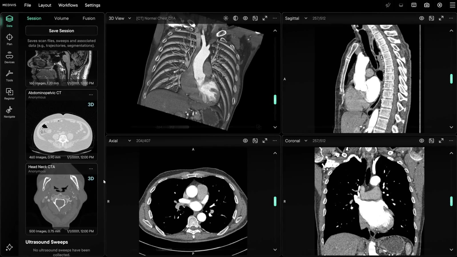
Session
Load from PACS, O-Arm, C-Arm, USB or CD into one session, saved once and recovered whole.
From the scan to the operating room. Medivis Studio turns imaging into a plan. Medivis Navigation carries that plan to the patient as real-time guidance. Frontier closes the loop, with systems that learn from every case.
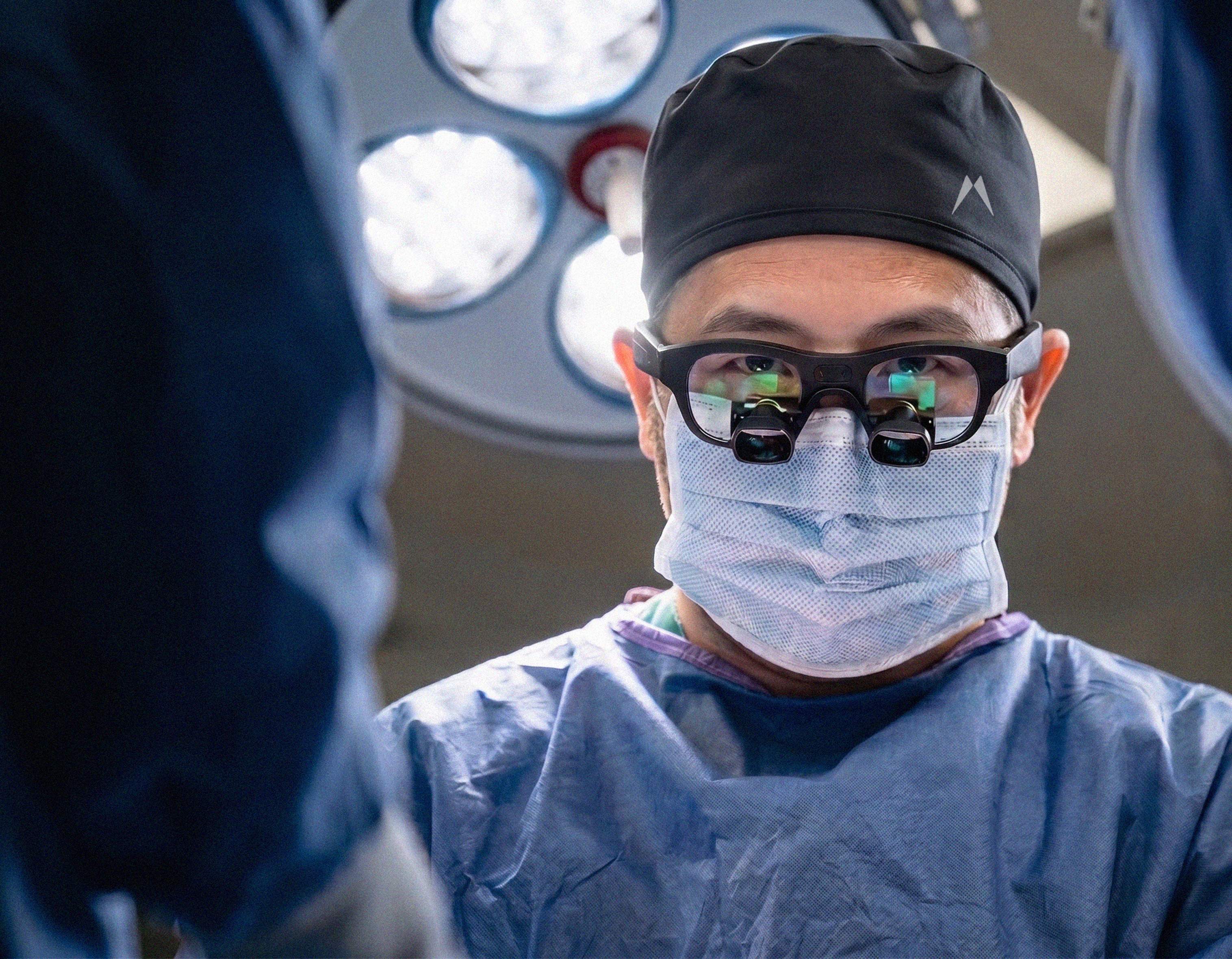
Smart Loupes are here
Spine Navigation →The FDA-cleared, CE-marked imaging platform for everything pre-operative — every case begins with the data, becomes a plan, and integrates with all the devices in the room. Available on Windows and macOS.
Everything a case is made of — CT, MRI, Ultrasound, Fluoroscopy, PET — the session it lives in, the volumes inside it, and the fusion of one modality with another.
Data
Load from PACS, O-Arm, C-Arm, USB or CD into one session, saved once and recovered whole.
Everything a case is made of — CT, MRI, Ultrasound, Fluoroscopy, PET — the session it lives in, the volumes inside it, and the fusion of one modality with another.
Load from PACS, O-Arm, C-Arm, USB or CD into one session, saved once and recovered whole.
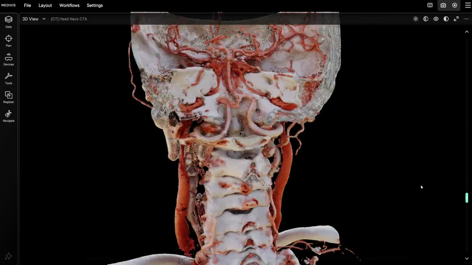
The 3D dataset cropped, cut and lit in real time, with photorealistic rendering on demand.
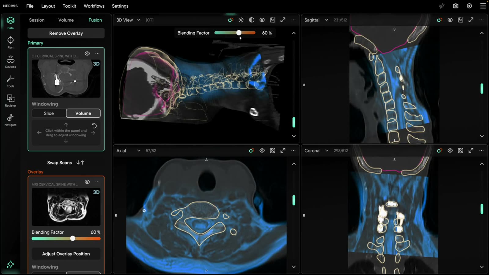
MRI fused onto CT with automatic registration; tune opacity and tint, or recompute any time.
Imaging becomes anatomy, and anatomy becomes a plan — segmented, annotated and templated on the patient’s own scan.
Plan
Automatic segmentation of spine, brain tumour and full-body CT, from a few seed points.
Imaging becomes anatomy, and anatomy becomes a plan — segmented, annotated and templated on the patient’s own scan.
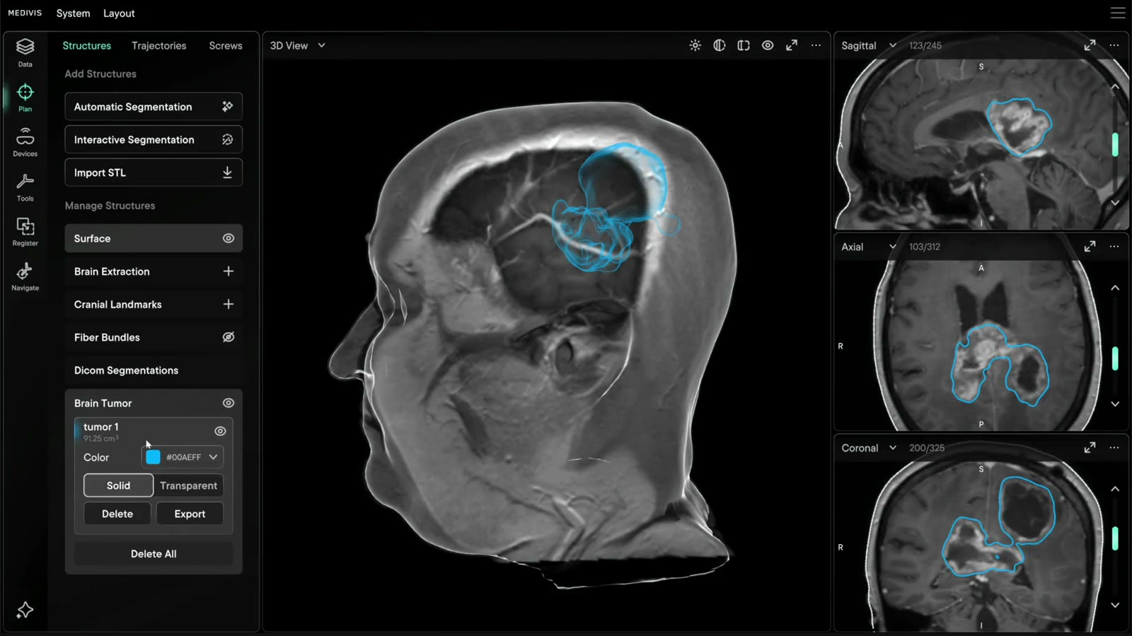
Automatic segmentation of spine, brain tumour and full-body CT, from a few seed points.
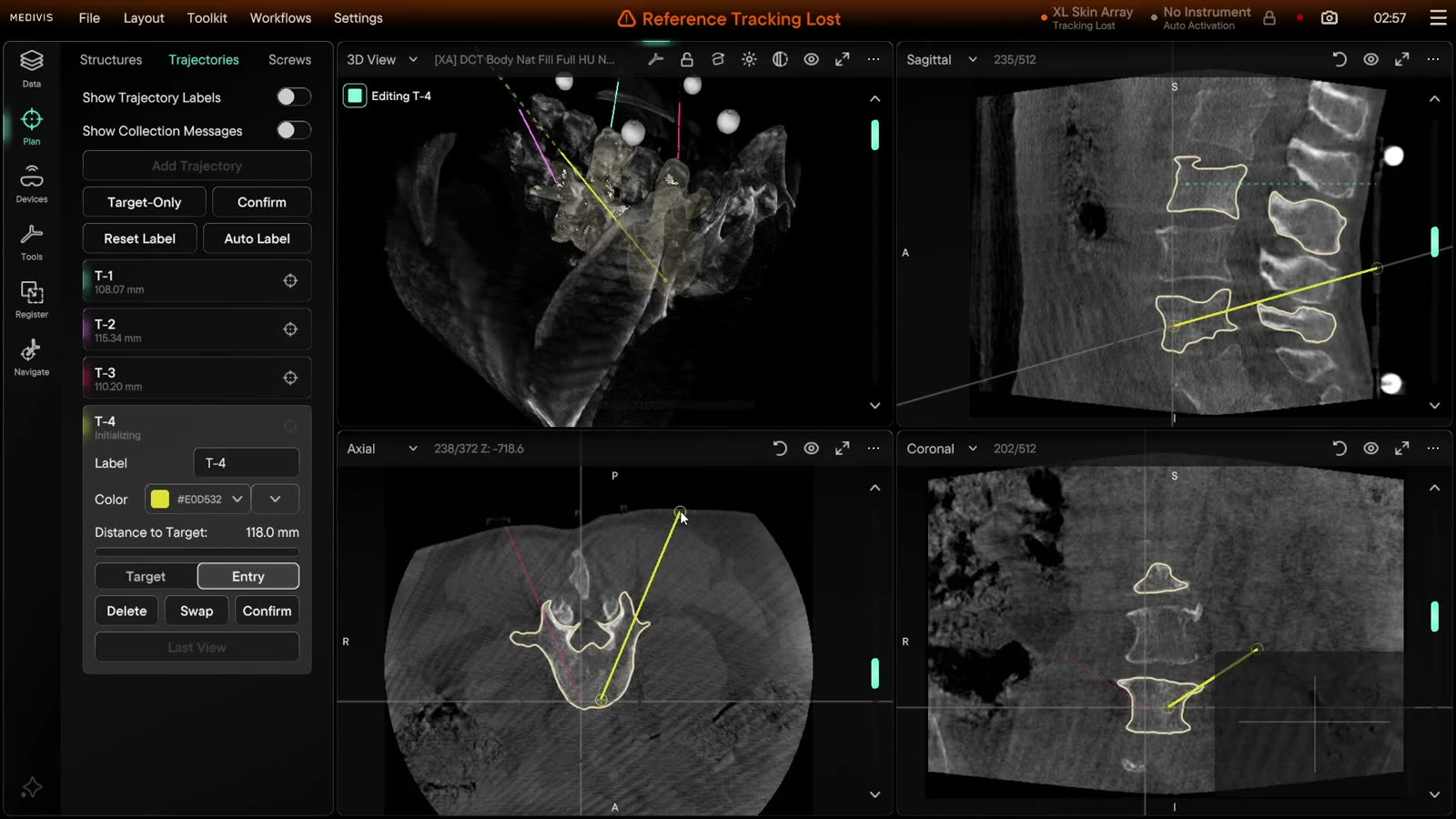
Target and entry trajectories on the patient’s own scan, measured to the millimetre.
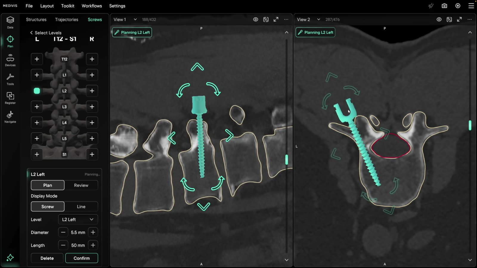
Screw planning level by level: diameter, length and trajectory, editable live in the OR.
The hardware around the case — sensors, headsets, screens, feeds and probes — connected to the same session so the room can see the plan.
Devices
Proprietary AR loupes, or commercial head-mounted displays from Meta, Apple and Microsoft. One session, any display.
The hardware around the case — sensors, headsets, screens, feeds and probes — connected to the same session so the room can see the plan.
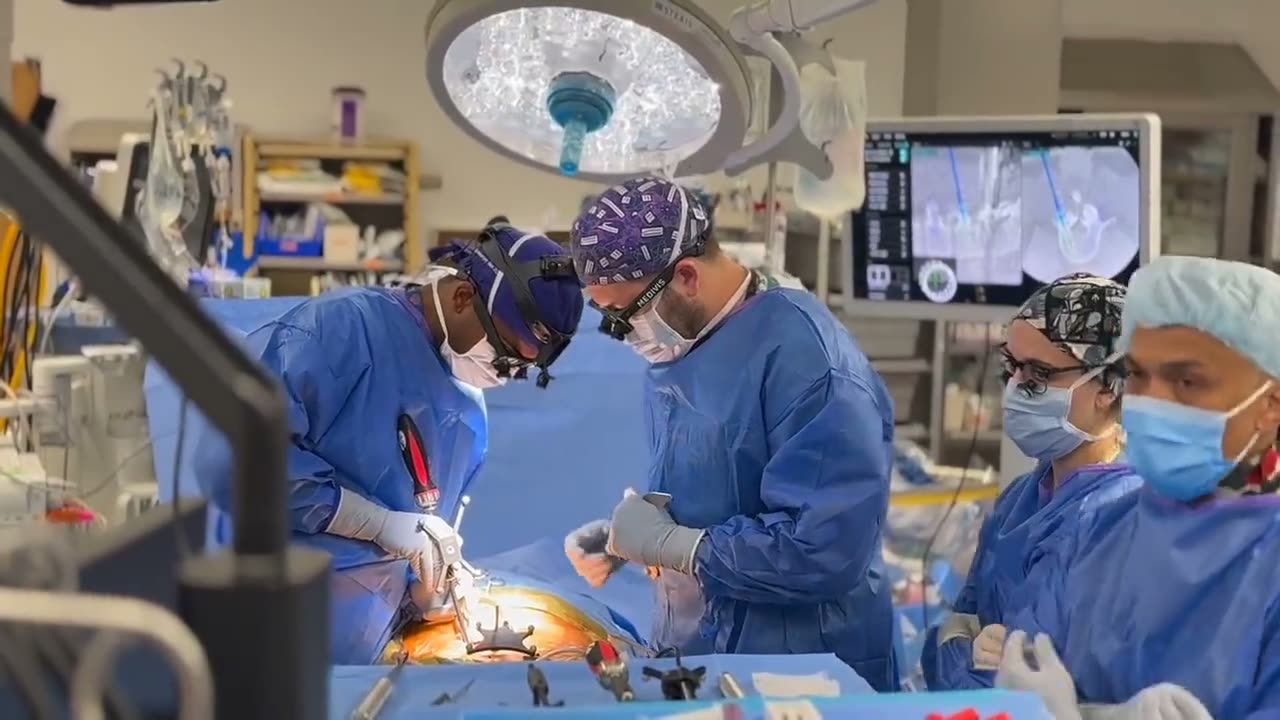
Proprietary AR loupes, or commercial head-mounted displays from Meta, Apple and Microsoft. One session, any display.
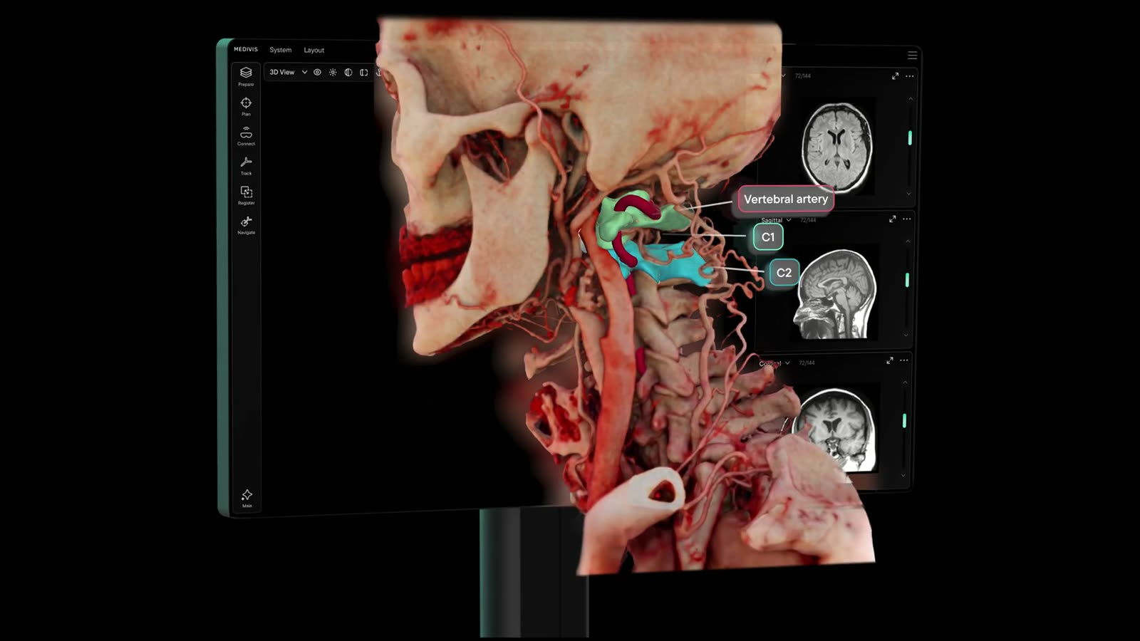
Glasses-free, eye-tracked 3D on the spatial monitor. No headset required.
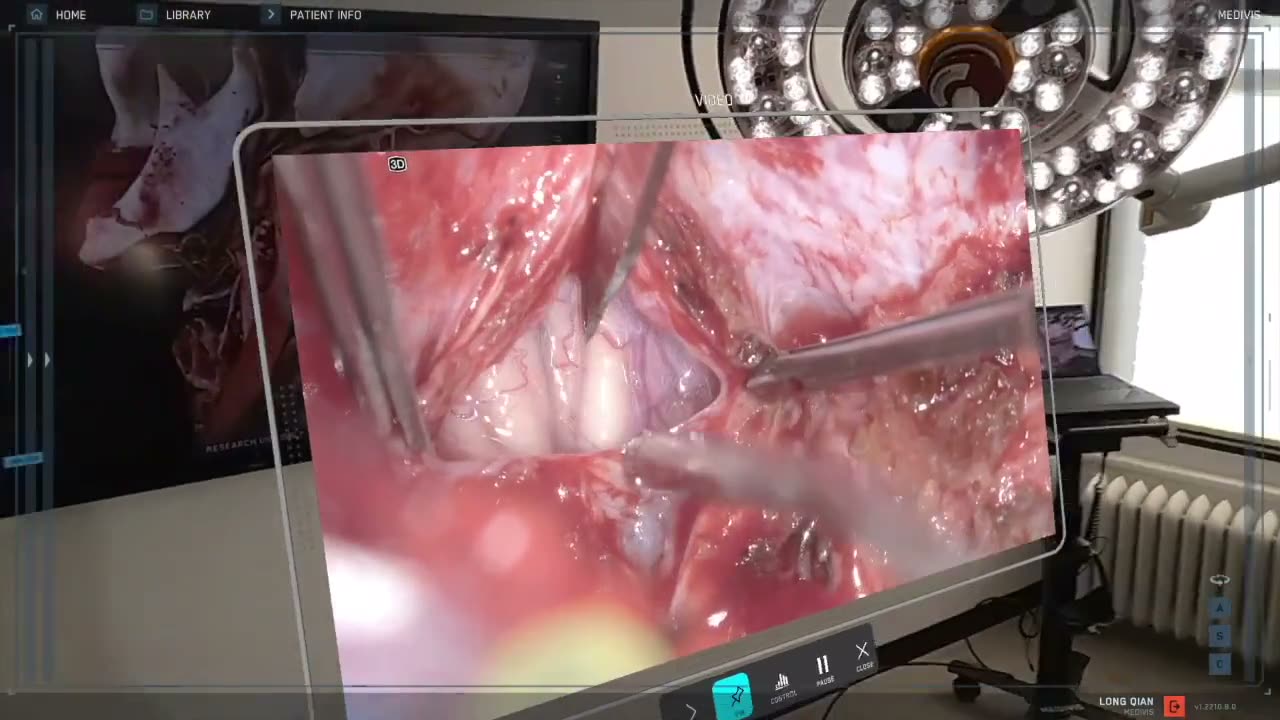
Cameras, endoscopes and microscopes routed into any viewport, or straight into the headset.
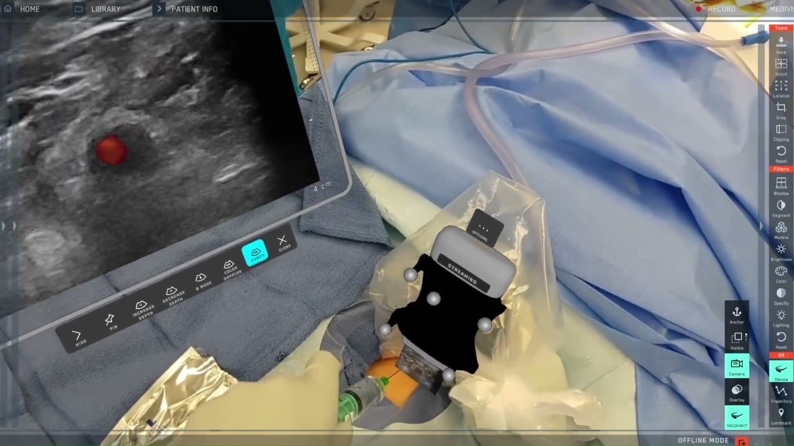
Tracked ultrasound in AR: B-mode and Doppler at the probe tip or on a virtual monitor.
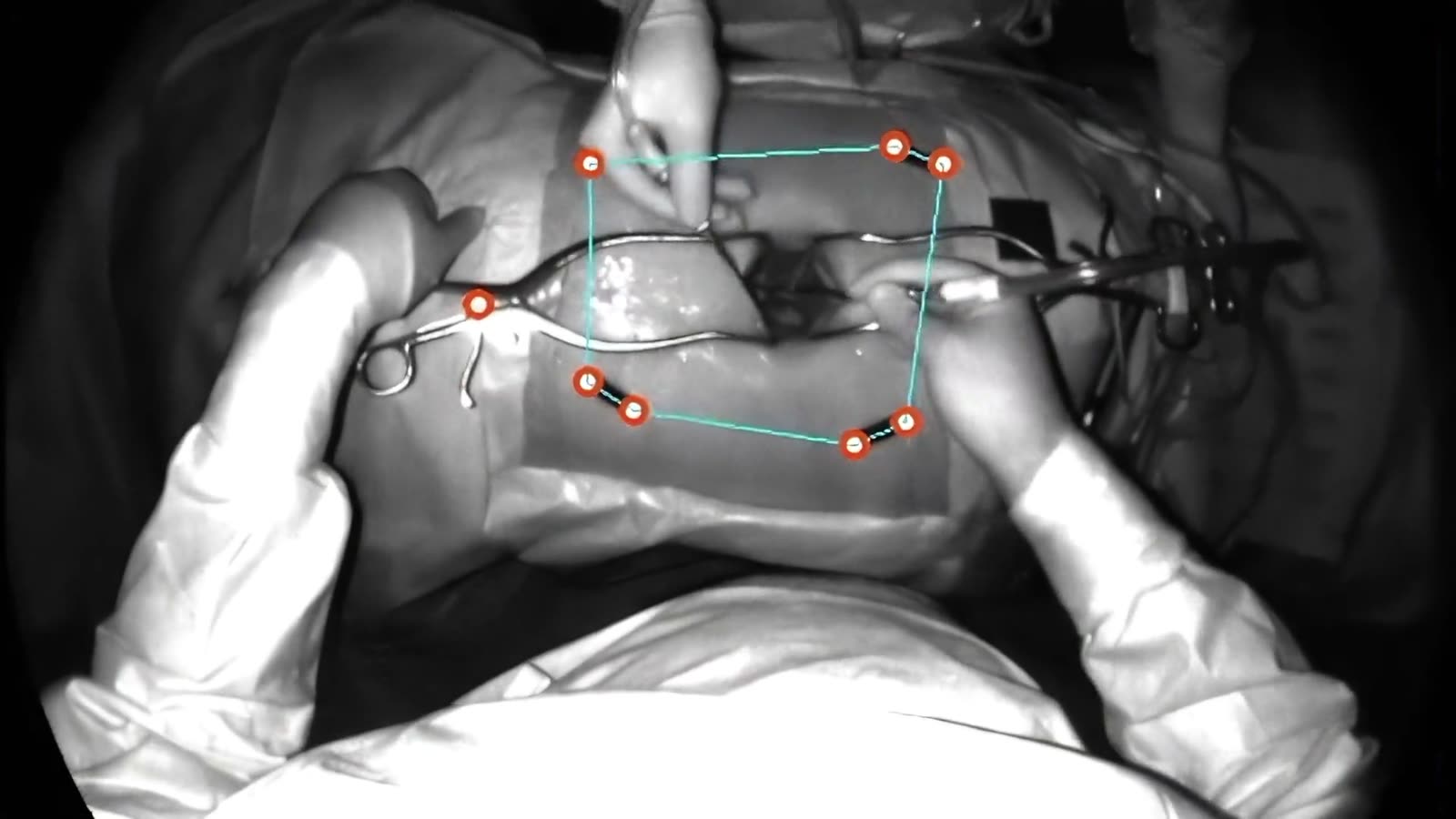
The optical tracker that sees the tools and the patient, with hybrid tracking through the headset.
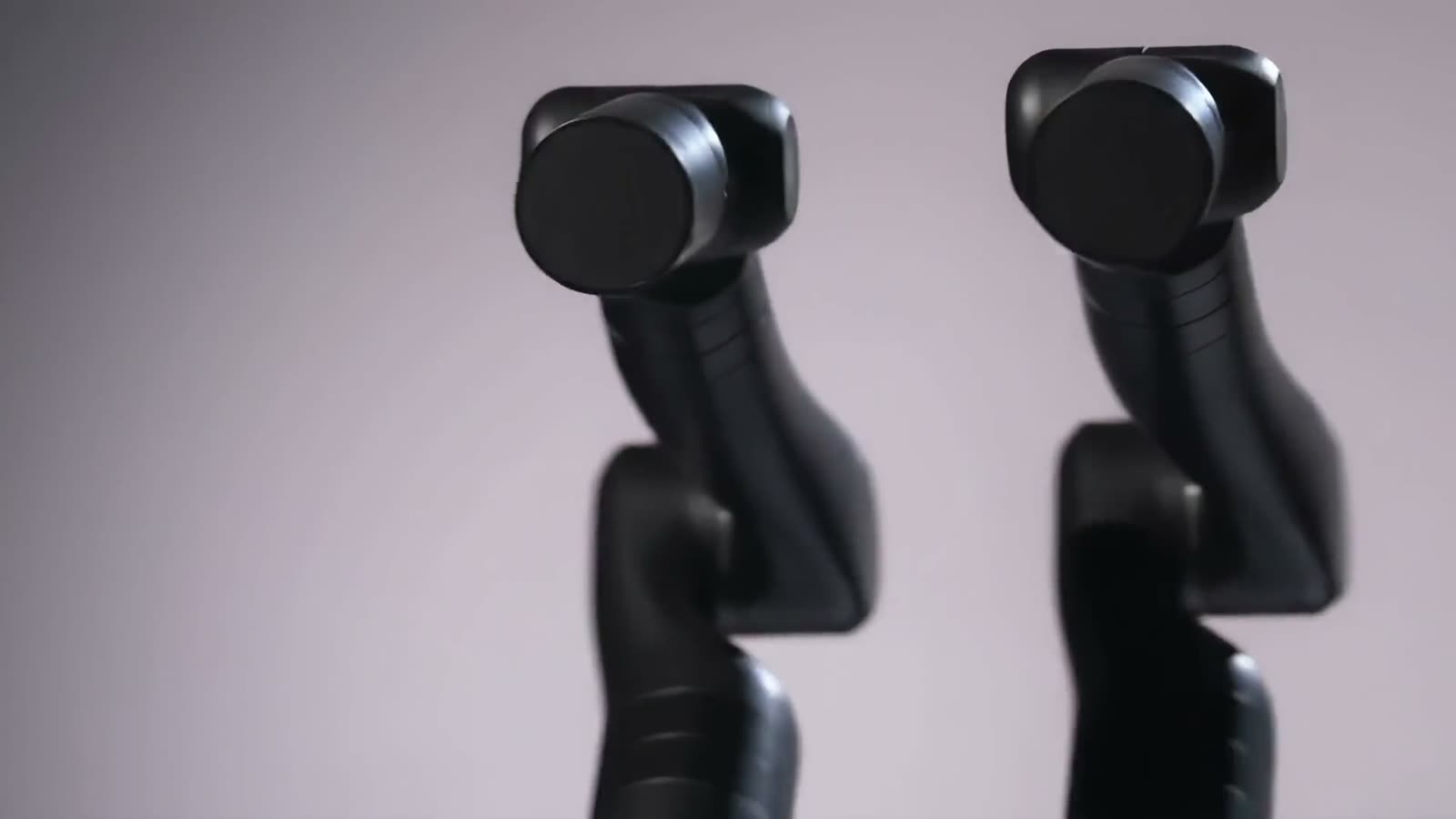
Tracked guidance extended to robotic positioning. Talk to us about integration.
Two ways to deploy
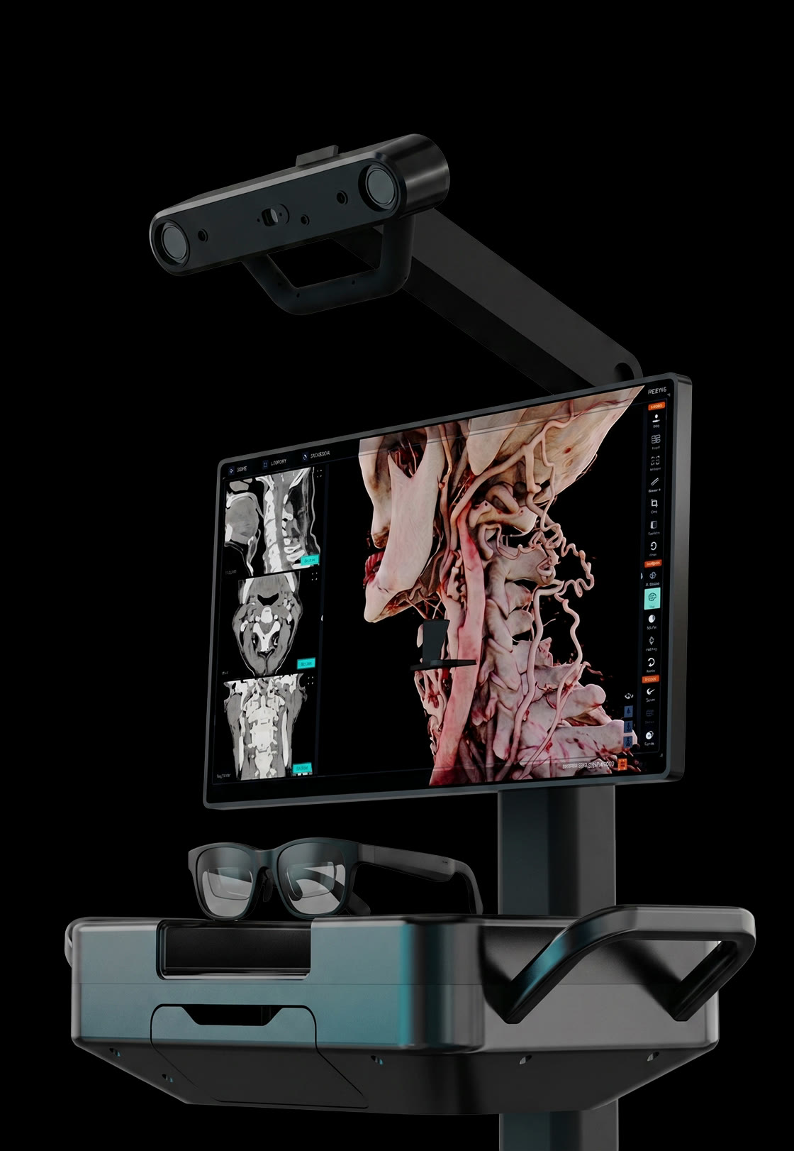
Execute the plan in the operating room with real-time guidance — 1st-party and 3rd-party tools, six ways to register, and conventional and/or spatial execution.
An ever-growing toolkit of 1st-party and 3rd-party instruments, references and adapters.
Tools
One library of trackable tools — both first-party and third-party versatility.
An ever-growing toolkit of 1st-party and 3rd-party instruments, references and adapters.
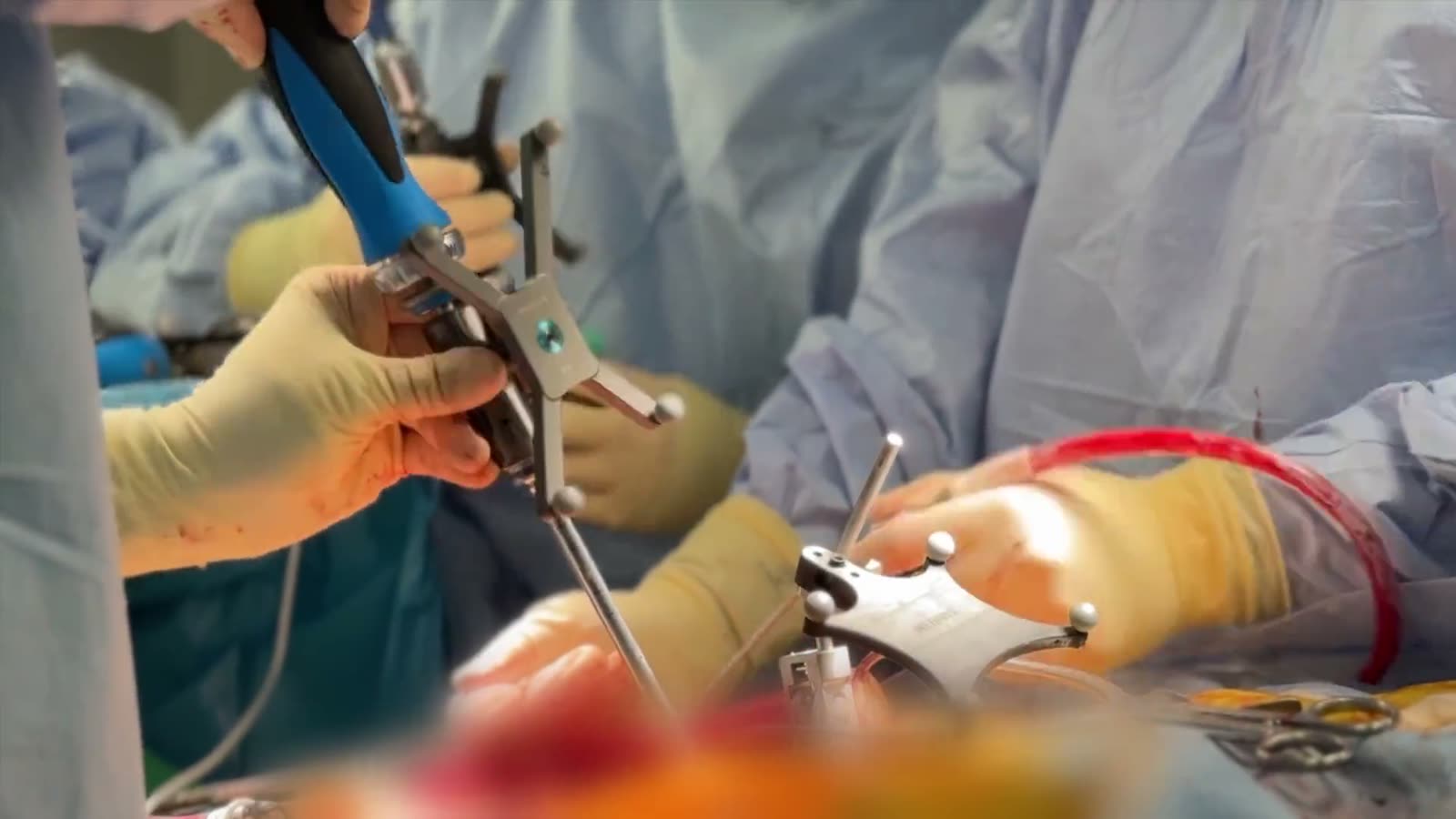
One library of trackable tools — both first-party and third-party versatility.
The moment the scan is locked to the patient — six ways to do it.
Register
Sparsely sample external or internal anatomy with a tracked tool, matched against the imaging isosurface, then registered.
The moment the scan is locked to the patient — six ways to do it.
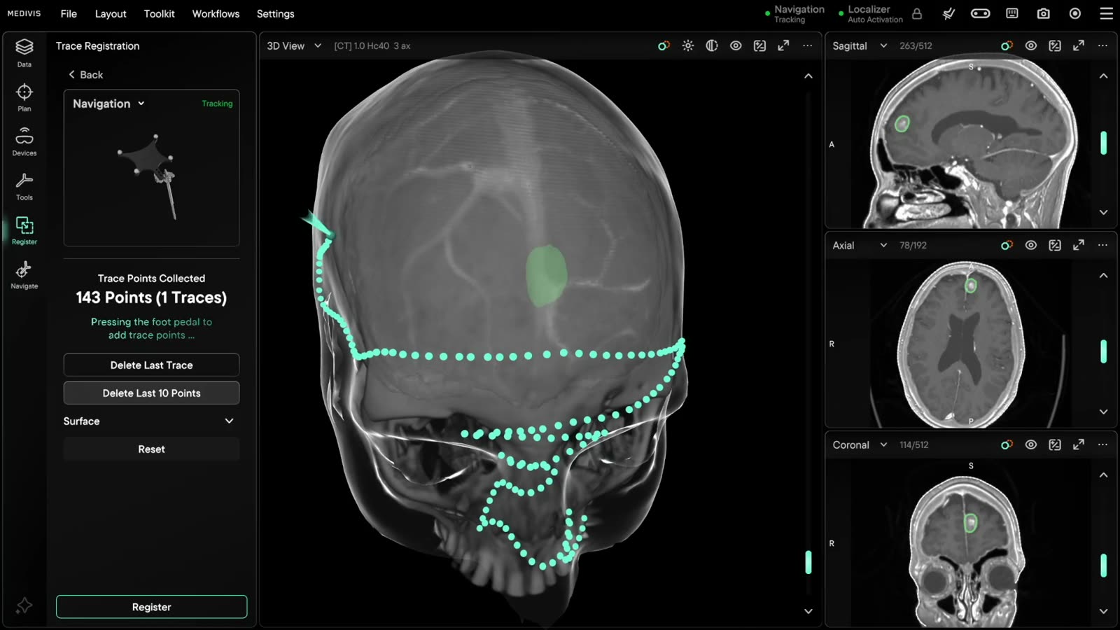
Sparsely sample external or internal anatomy with a tracked tool, matched against the imaging isosurface, then registered.
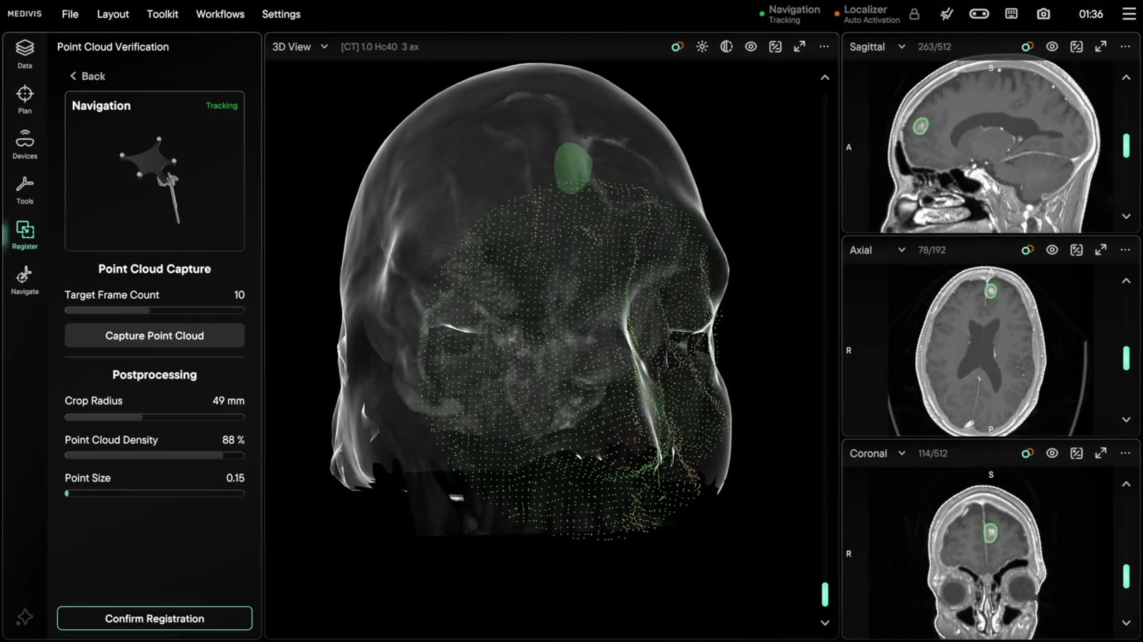
Surface digitization with high-density point cloud captures, verified against the scan, automatically, in seconds.
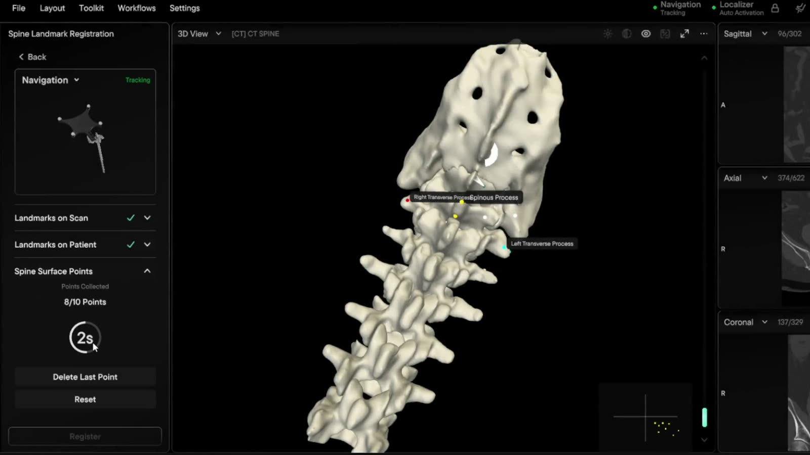
Virtual landmarks on the scan, the same points on the patient — the spatial alignment is instantly computed.
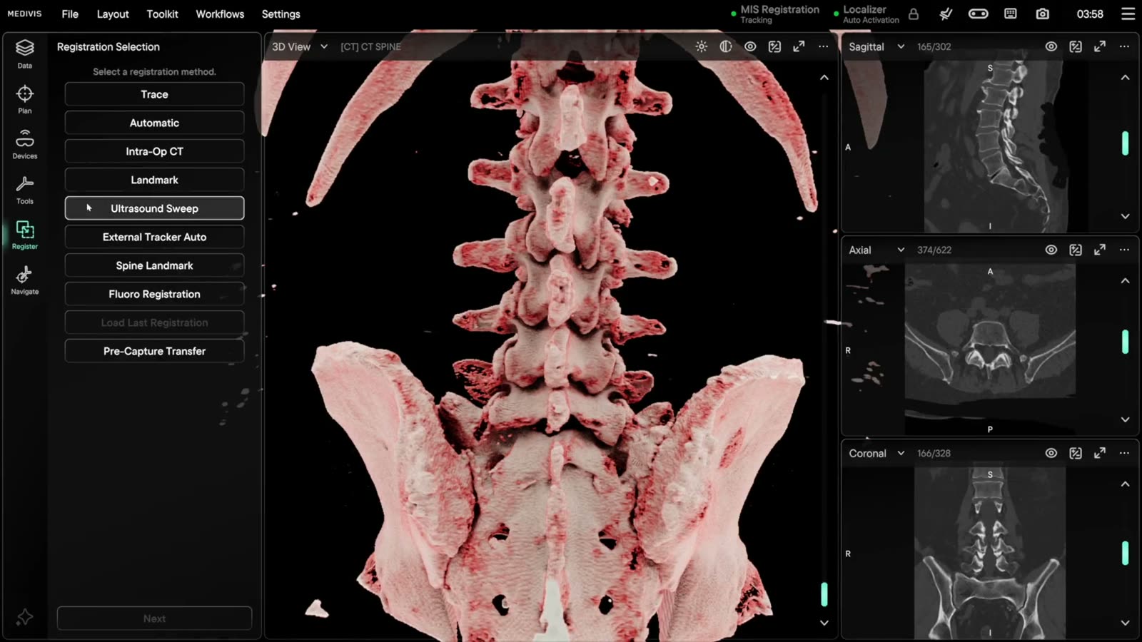
Registration from the scan itself: fiducials found automatically and aligned to the array.
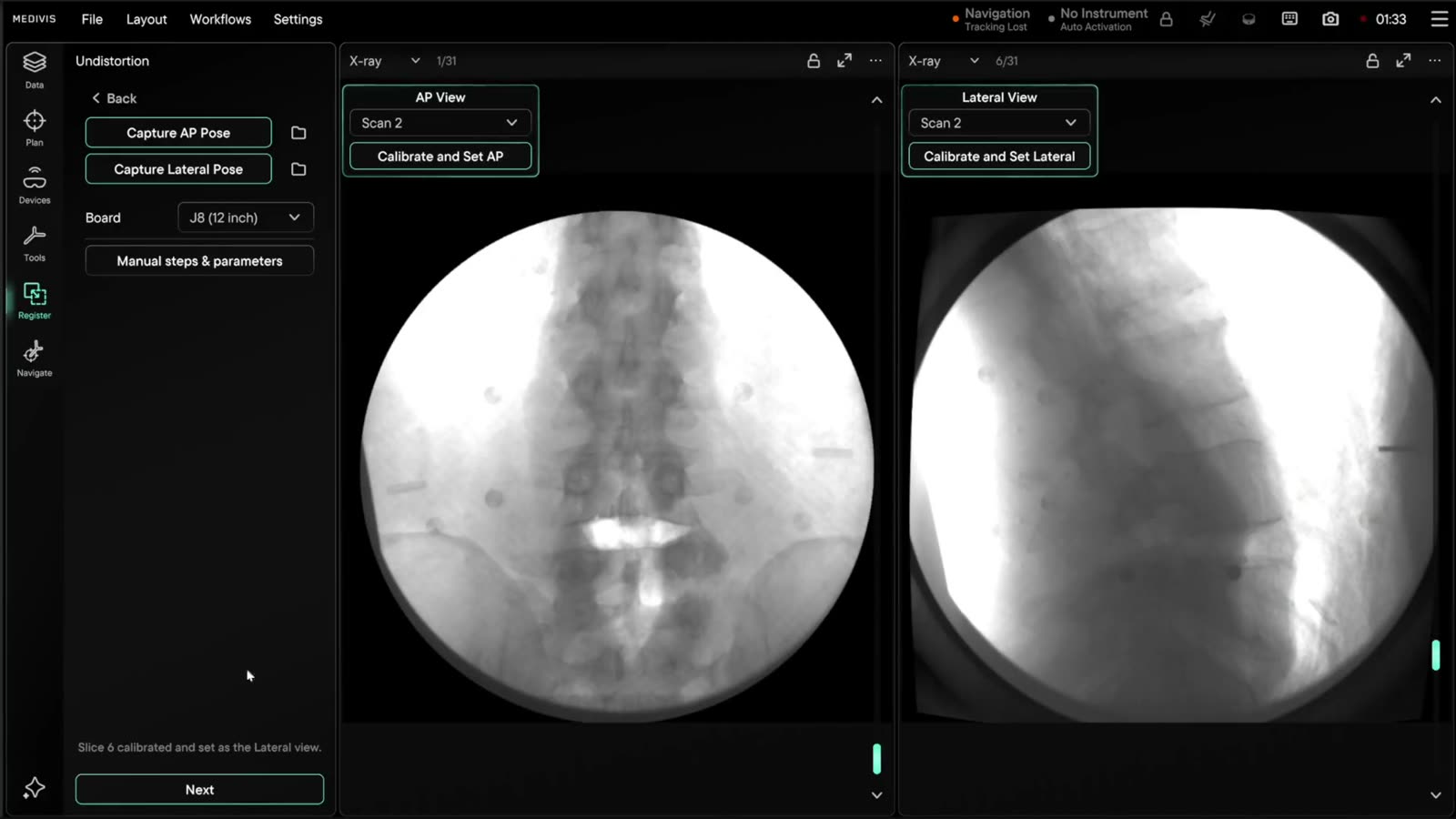
Fluoroscopy registered to the volume. Talk to us about your program.
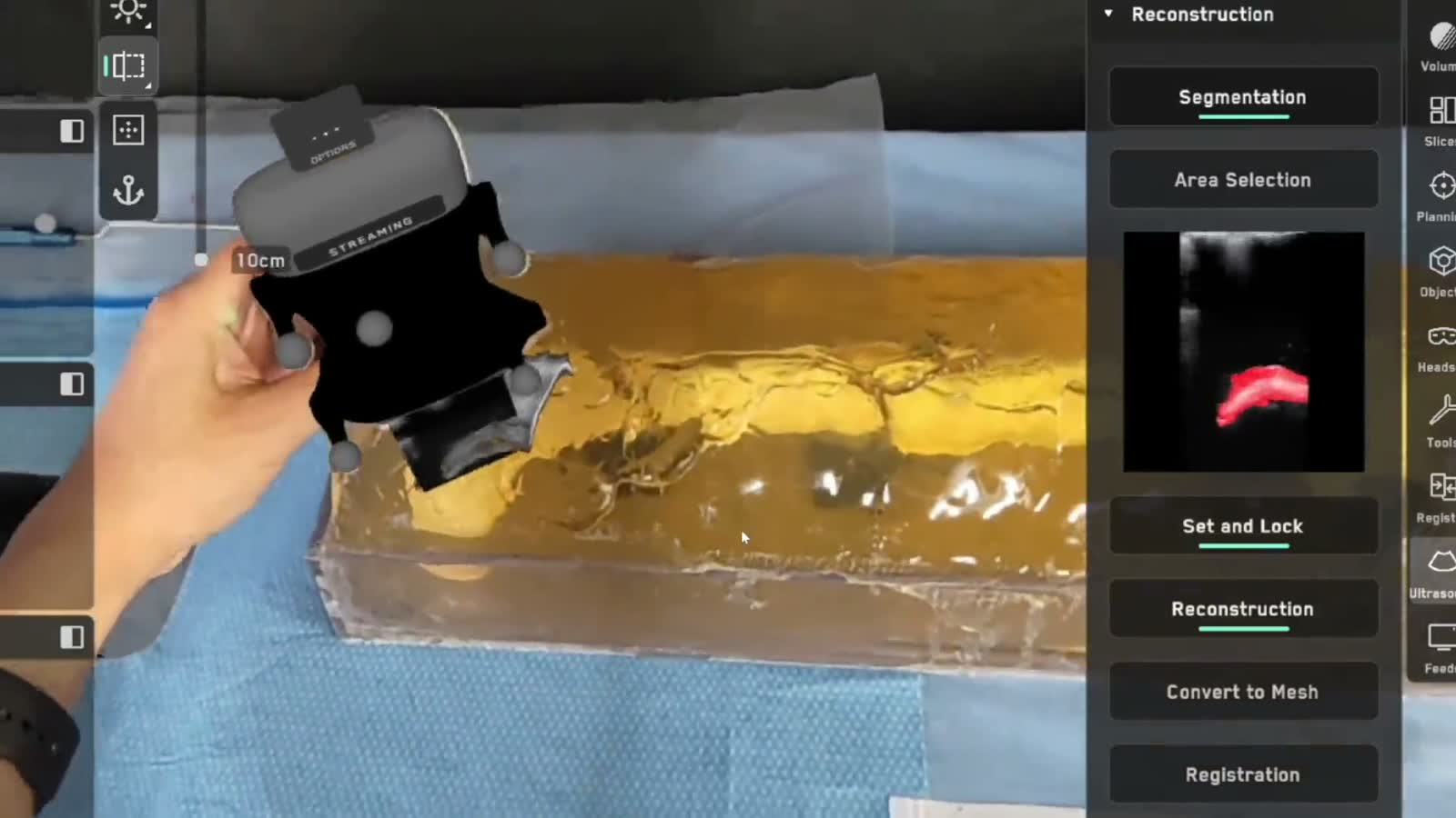
A sweep of the tracked probe digitizes internal anatomy in seconds, which can be fused with cross-sectional imaging.
Conventional and/or spatial execution. Cranial and spine FDA cleared. Body under investigation.
Navigate
FDA cleared for use in the OR, ICU or bedside — from craniotomies to EVDs.
Conventional and/or spatial execution. Cranial and spine FDA cleared. Body under investigation.
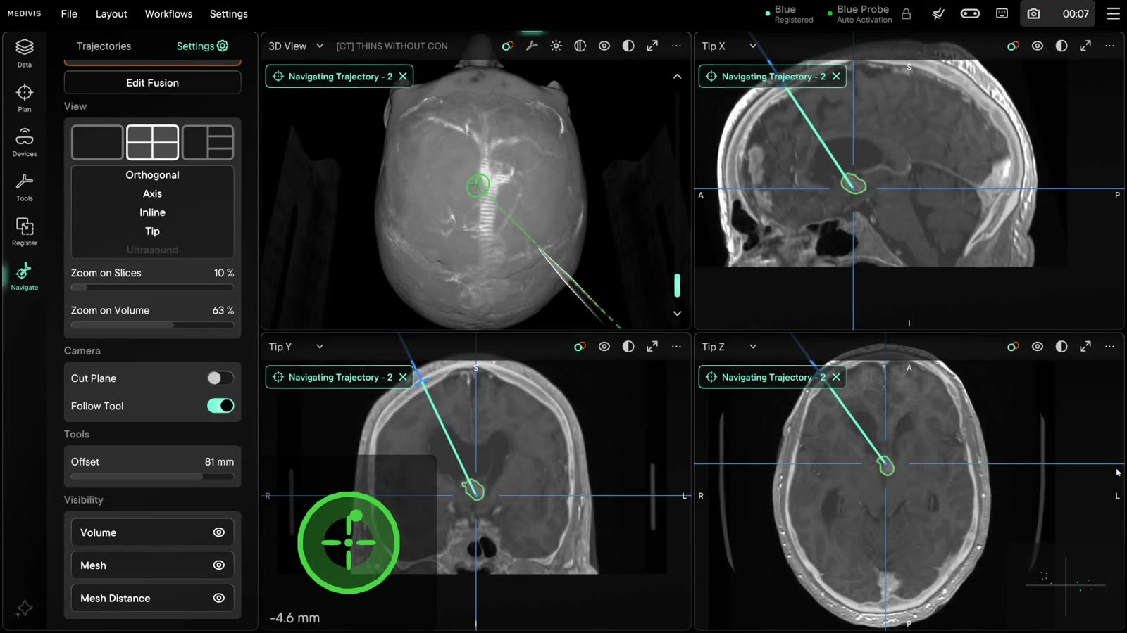
FDA cleared for use in the OR, ICU or bedside — from craniotomies to EVDs.
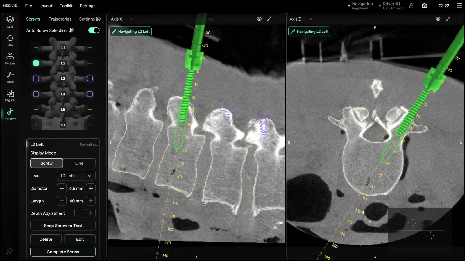
FDA cleared for thoracolumbar spine, open and minimally invasive. Cervical spine navigation: FDA clearance pending.
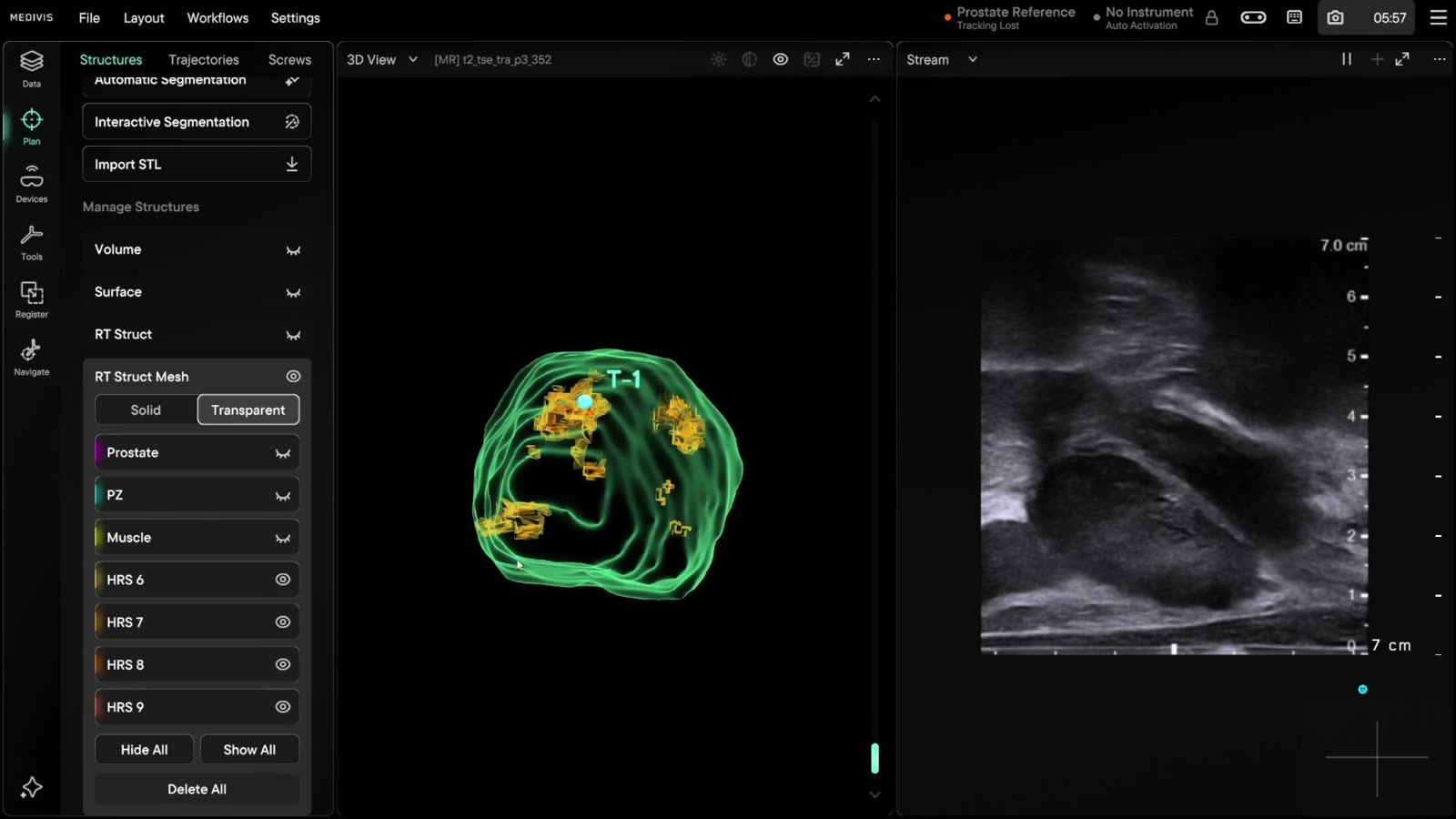
Investigational — FDA clearance pending. Real-time ultrasound superimposed on the patient, fused with MRI.
Where our research is heading — an agent that is really a contextual harness, and robots that join a system which already knows where everything is.
Frontier: co-development programs, not cleared products.
Frontier
Maia puts the imaging and live case state in front of any model, then calls our validated tools.
Where our research is heading — an agent that is really a contextual harness, and robots that join a system which already knows where everything is.
Frontier: co-development programs, not cleared products.
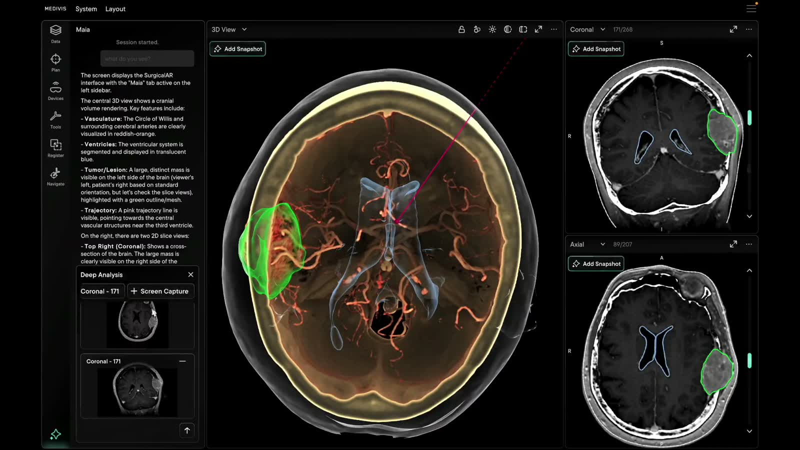
Maia puts the imaging and live case state in front of any model, then calls our validated tools.
Surgical autonomy runs 0 to 5. The field has cleared Level 2; above it is our research. Research use only.

Founded by physicians and engineers in New York. We build the whole loop, from the scanner to the operating room, so the surgeon never has to look away from the patient.
Send us a message — a member of the Medivis team replies within one business day.
